Case of the Month: Bilateral Testicular Swellings in a Young Male
Case Authors: Numbereye Numbere, MBBS, Hiroshi Miyamoto, MD, PhD
Clinical History
A young (3rd decade) male presents with a history of male infertility. Examination revealed bilateral painless testicular masses.
Past Medical History
The patient's history was notable for congenital adrenal hyperplasia on treatment. Scrotal ultrasound showed bilateral intratesticular masses with heterogeneous echotexture and vascularity. An excisional biopsy was performed.
Recent History
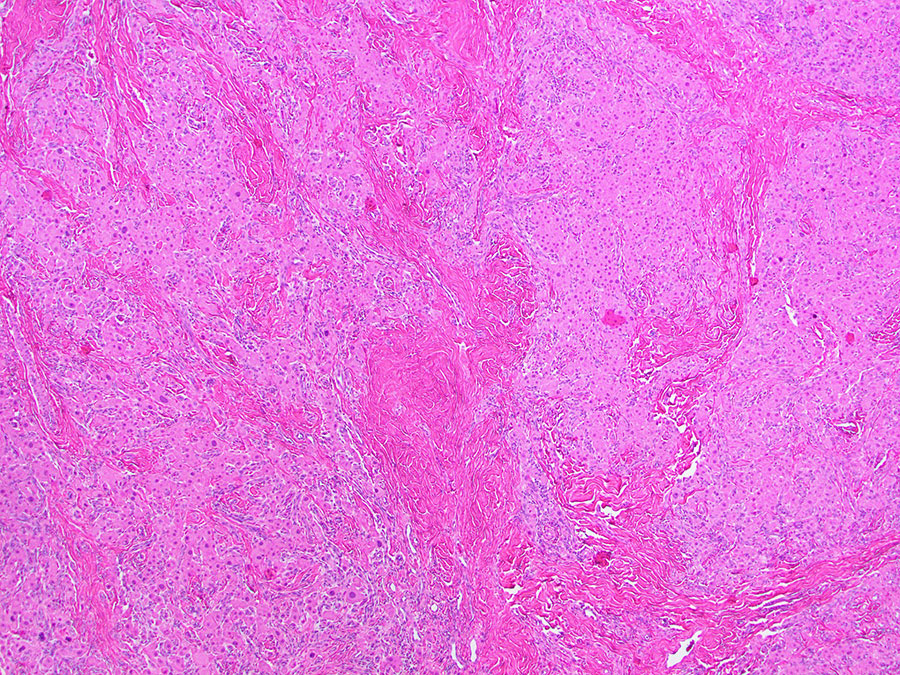
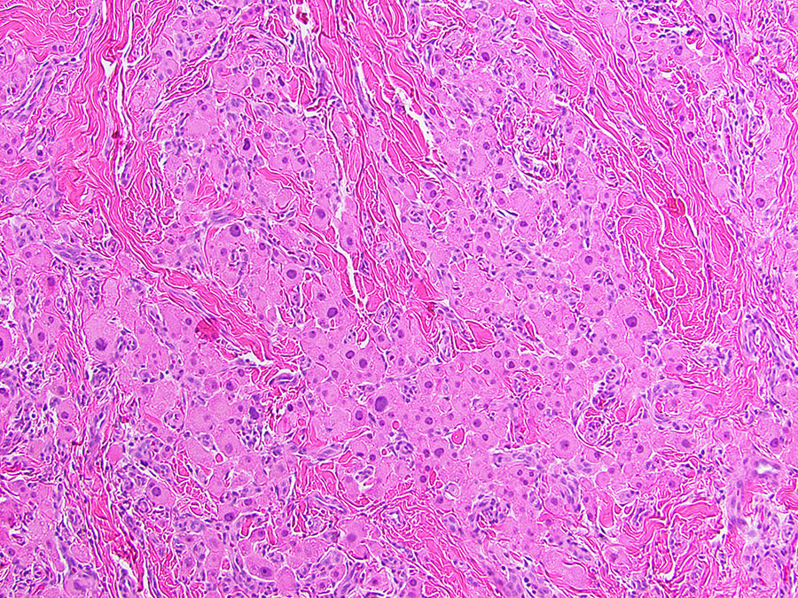
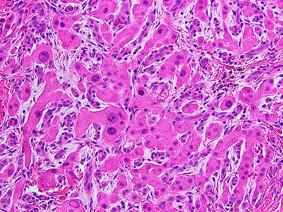
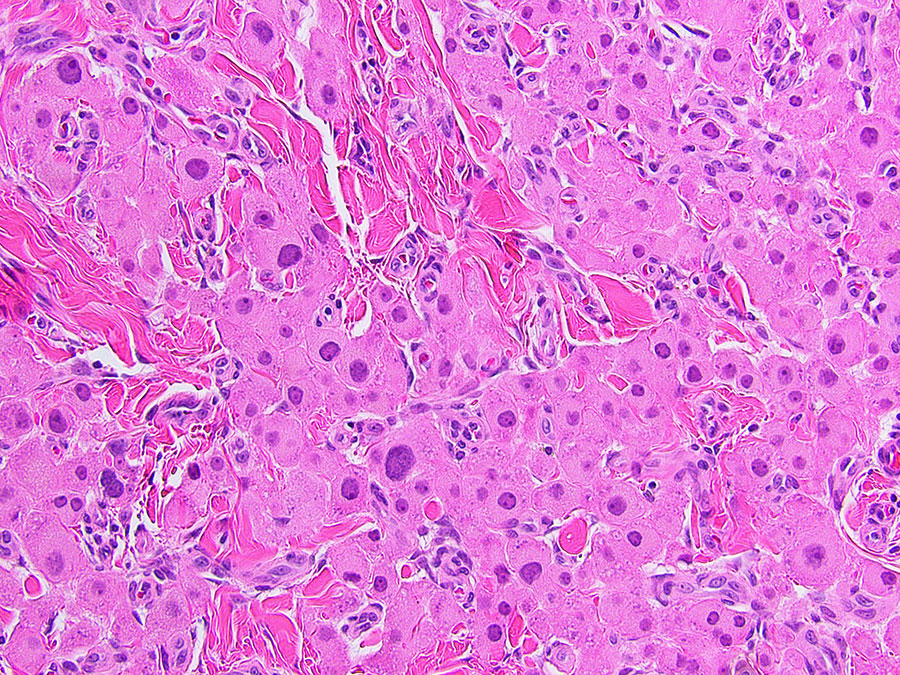
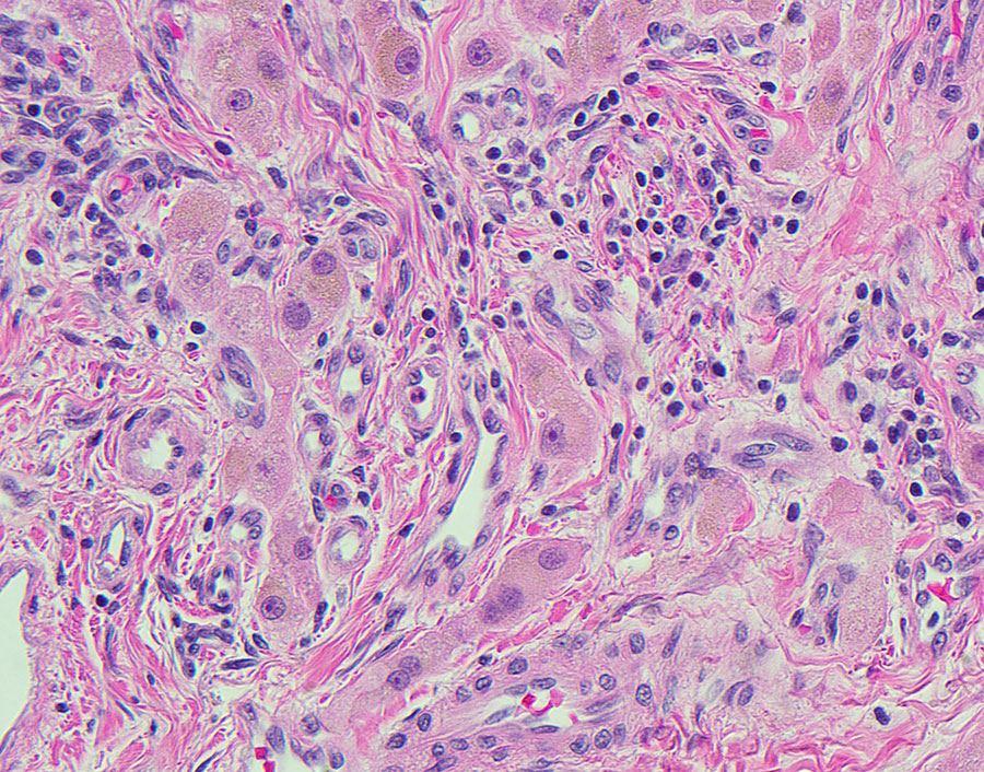
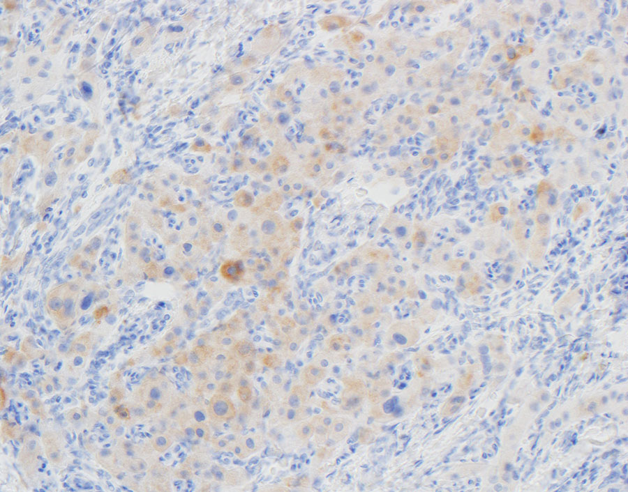
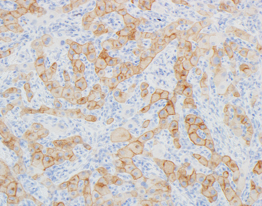
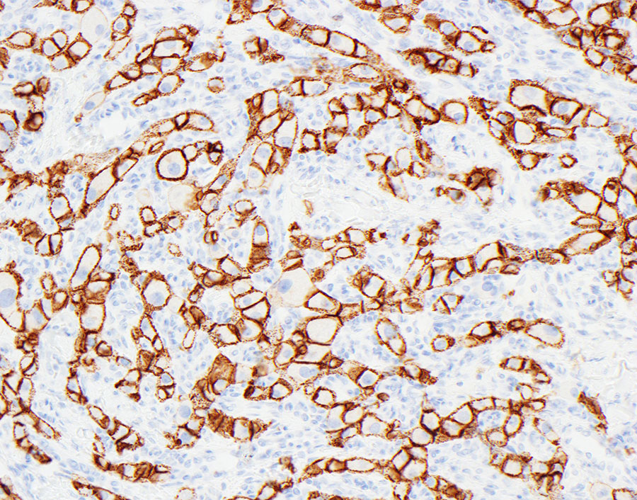
At surgery, there were multiple hard testicular masses with compression of adjacent testicular tissue and dilatation of upstream seminiferous tubules. Microscopic examination of the excised masses showed sheets, cords, and lobules of large, polygonal cells with abundant, finely granular eosinophilic cytoplasm and large pleomorphic nuclei (Figures 1-4). Intracytoplasmic lipochrome pigment was seen in areas (Figure 5). The lesional cells were positive for inhibin (Figure 6), synaptophysin (Figure 7), and CD56 (Figure 8) but negative for androgen receptor (not shown). Adjacent uninvolved testis was unremarkable and showed preserved spermatogenesis.