Case of the Month: Pancreatic Mass with Metastases to the Liver
Case Author: Numbereye Numbere, MBBS
Clinical History
An elderly man has a history of a 6.3 cm. mass in the body and neck of the pancreas.
Past Medical History
The patient presented four weeks earlier with epigastric pain and bloating. The patient's medical history is notable for a remote history of completely resected colonic adenocarcinoma without recurrence.
Recent History
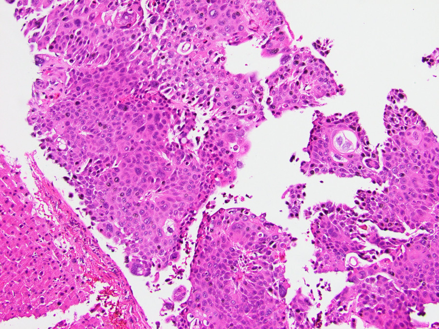
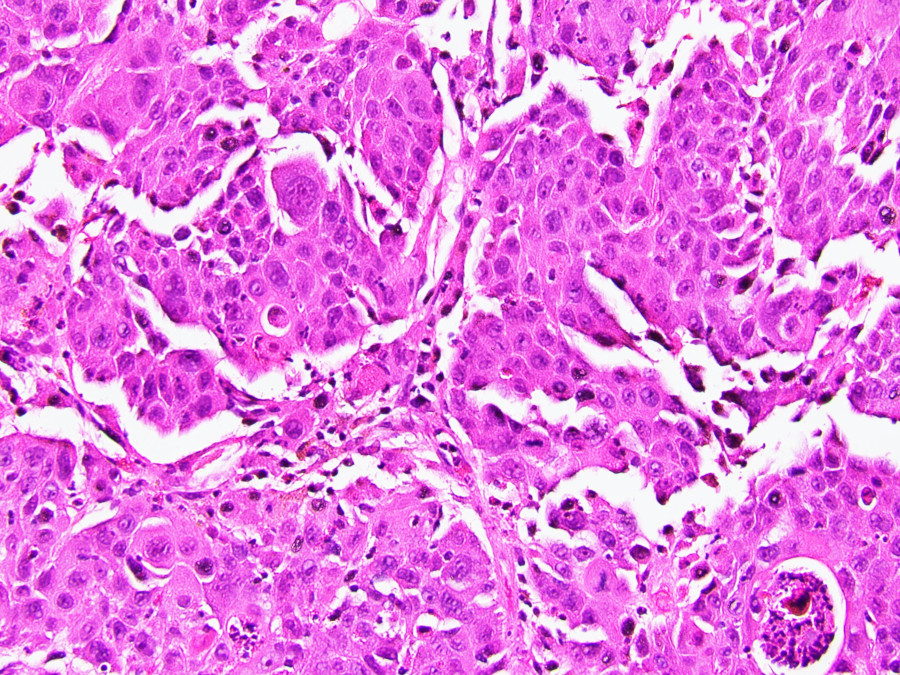
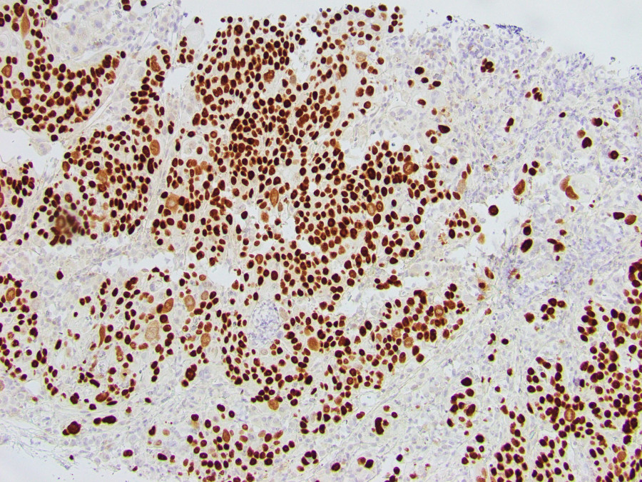
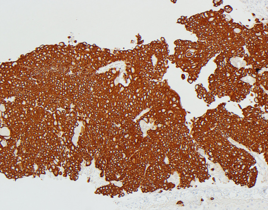
Follow-up ultrasound scan and magnetic resonance imaging revealed a 6.3 cm centrally necrotic mass in the body and neck of the pancreas with metastatic lesions in the liver. Percutaneous pancreatic and liver biopsies were performed. Histologic evaluation showed sheets of atypical cells with abundant eosinophilic cytoplasm, prominent intercellular bridges, and apparent keratin (figures 1 and 2). Immunohistochemical stains for p40 (figure 3) and cytokeratin 5/6 (figure 4) were strongly positive. A mucicarmine stain was negative.