Elderly Male with Rapidly Worsening Sinusitis
Talia Chen, MD, Alicia Schmidt, DO, Xiaolan Ou, MD, Numbereye Numbere, MD
Clinical History
An elderly male presented with 1 week of headaches, progressive vision loss in the right eye, and impaired extraocular movements.
Past Medical History
The patient has a history of chronic sinusitis status post endoscopic sinus surgery. Additionally, the patient was undergoing chemotherapy for advanced carcinoma.
Recent History
After surgery, the patient continued to experience purulent nasal drainage and headaches. Nasal cultures taken four weeks post-op grew only Stenotrophomonas and Achromobacter. He was treated with antibiotics, but his symptoms persisted. A week later, he experienced a darkening of vision in his right eye along with worsening headaches, prompting him to present at the ED.
Upon admission, the patient’s previous nasal cultures were found to have added growth of Aspergillus. Contrast-enhanced MRI and CT of the head and paranasal sinuses showed mucosal thickening of the bilateral sinus cavities, features consistent with osseous invasion, and leptomeningeal and cerebral involvement. The patient was treated with voriconazole and underwent extensive surgical debridement of the sinus cavities.
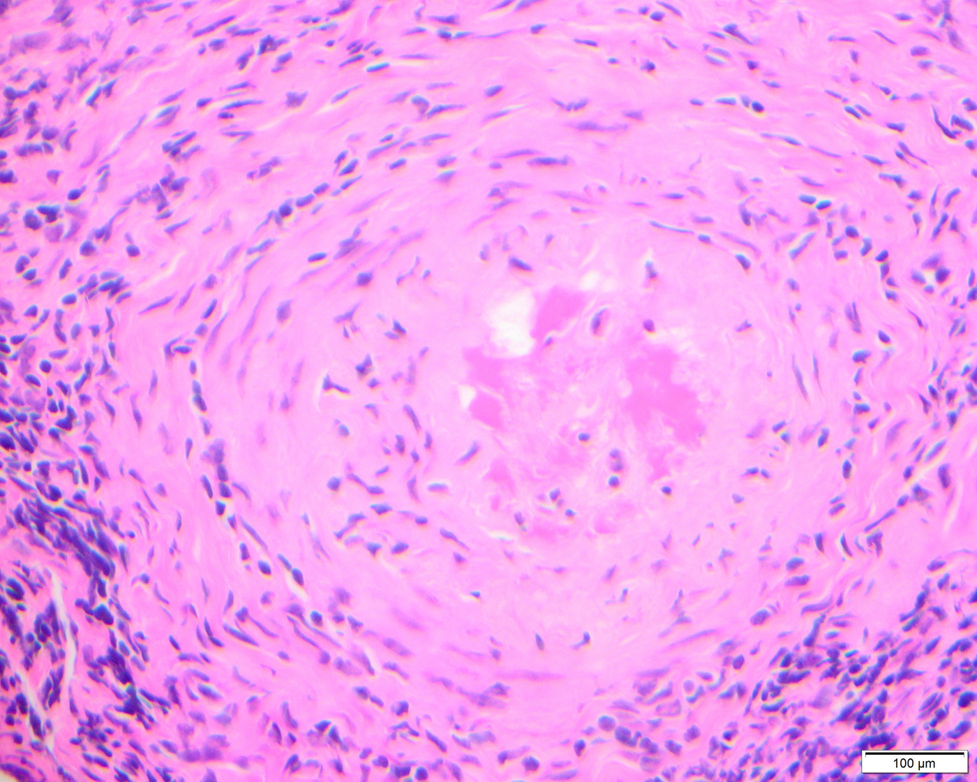
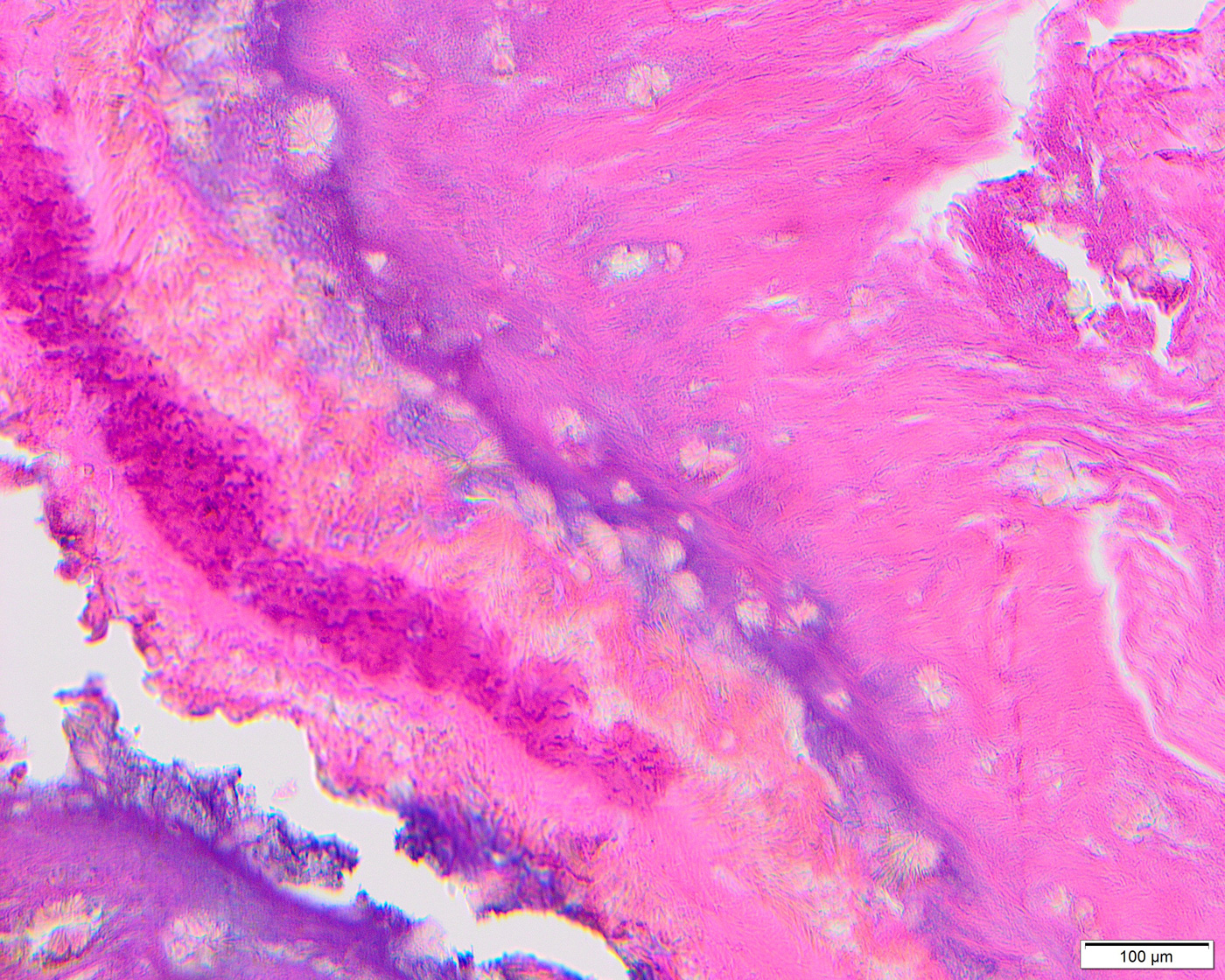
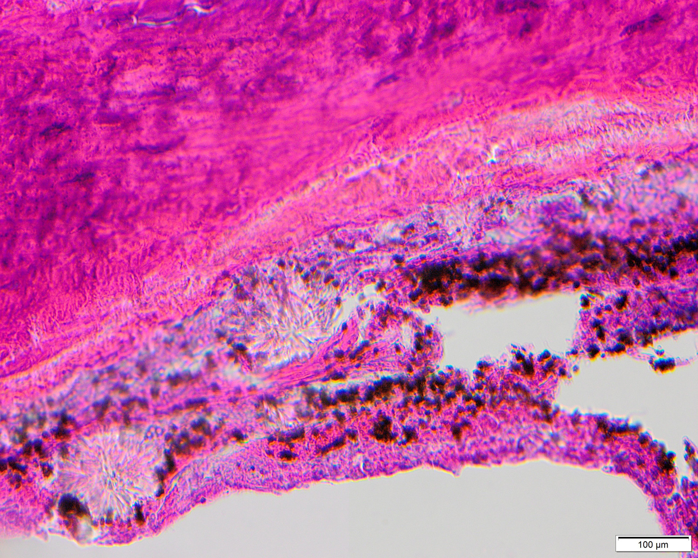
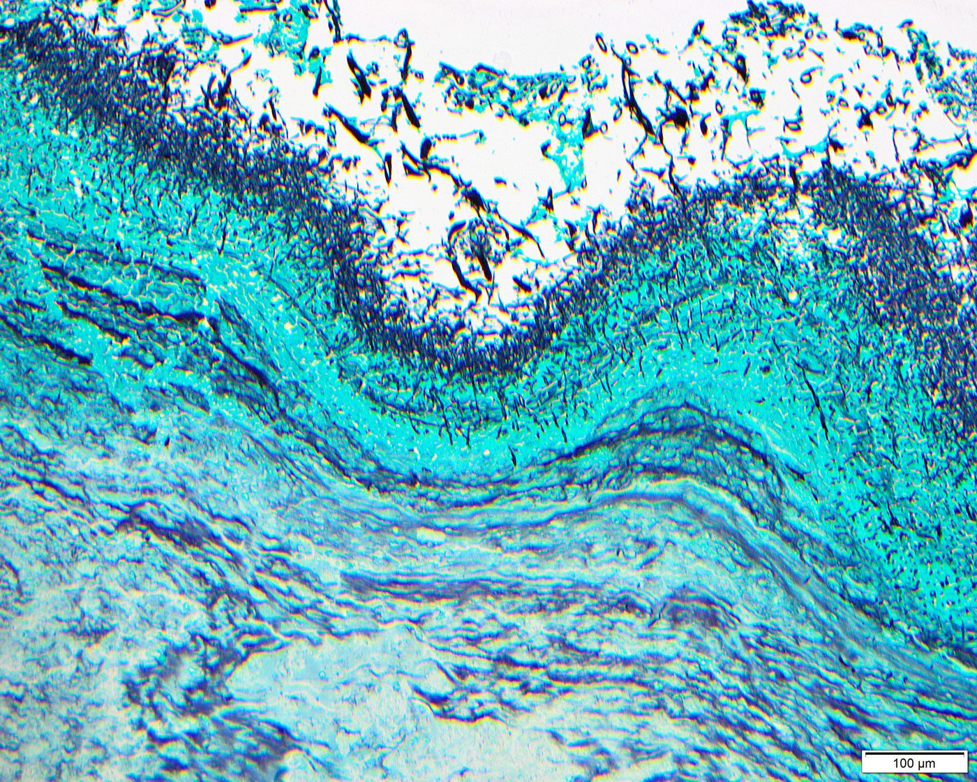
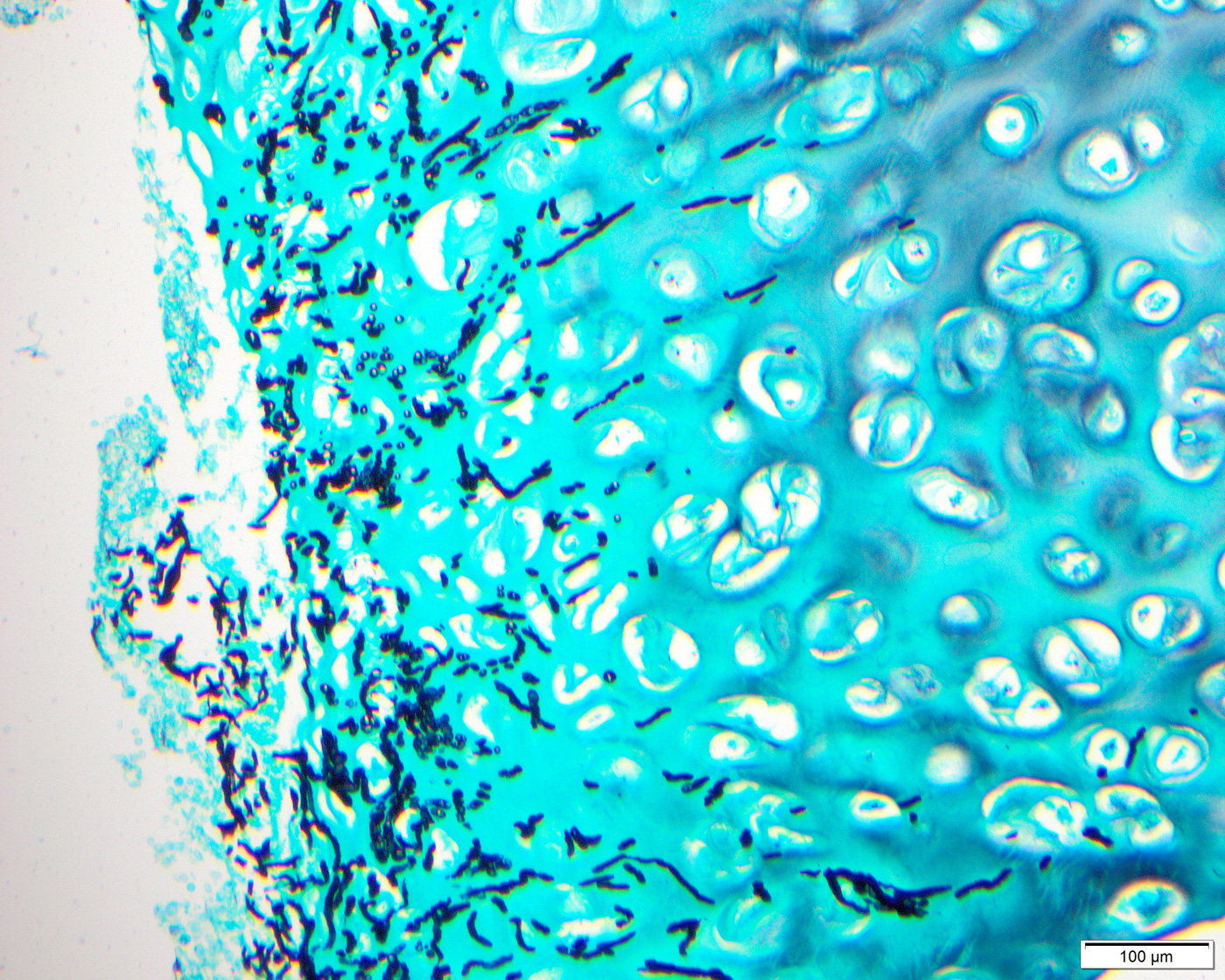
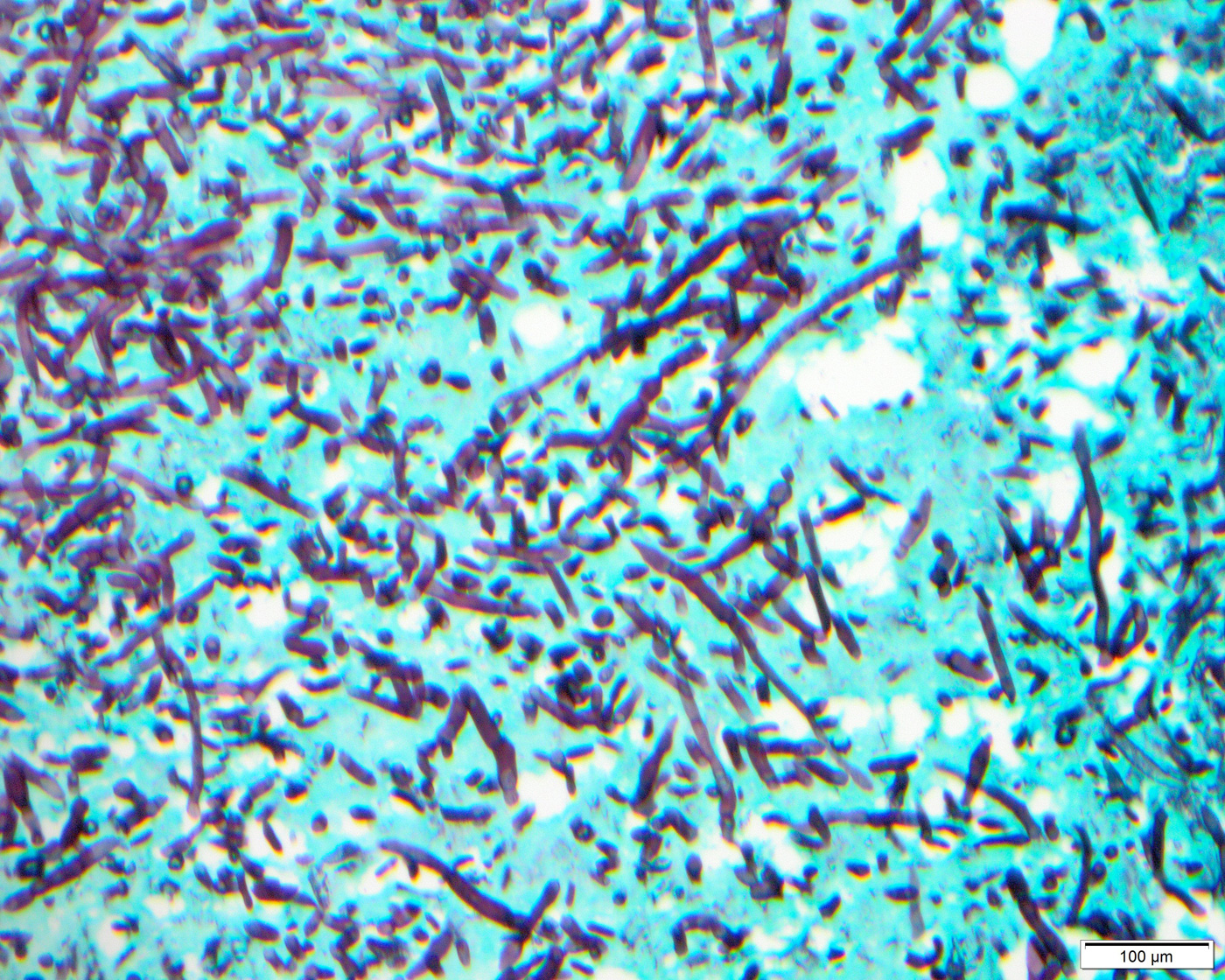
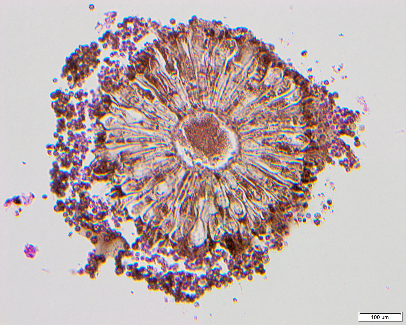
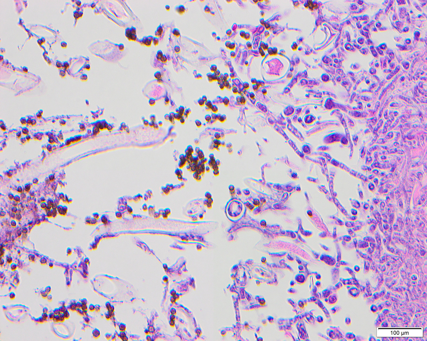
Histologic sections of the debrided tissue showed extensive necrosis of the sinonasal mucosa, soft tissue, and cartilage, abundant mixed inflammatory cells, and fungal organisms with features compatible with vascular invasion (Fig. 1). Intravascular thrombi (Fig. 1) and calcium oxalate crystals (Figs. 2 and 3) were also identified. The fungi were highlighted by GMS stain (Figs. 4 and 5) and displayed unpigmented septate acute-angle branching hyphae (Fig. 6) and fruiting bodies (fig. 7). Another fungal organism with wider hyphae, morphologically suggestive of Fusarium or Scedosporium was seen (Fig. 8). However, since no other fungi were isolated from culture, its exact identification remains unconfirmed.