Case of the Month: Enlarging Breast Mass
By Alison Gaylo, Ph.D., M.D. candidate
Clinical History
A 17-year-old female presented with an enlarging and increasingly tender left breast mass at the 7 o’clock position between the inframammary crease and the nipple areolar complex.
Past Medical History
No significant medical or surgical history.
Recent History
An elective resection was performed of the left breast mass. On excision, the mass was noted to be relatively superficial and well-circumscribed. A 9 x 7 x 3 cm ovoid mass was obtained, with an attached skin ellipse. Serial sectioning demonstrates predominately tan-pink, homogenous, dense and cystic fibrous tissue, with cysts being up to 0.5cm in diameter. There was no obvious necrosis, calcifications, or hemorrhage identified.
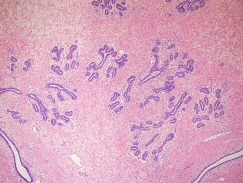
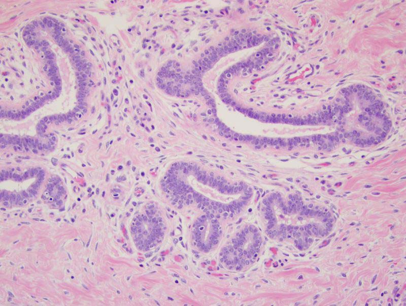
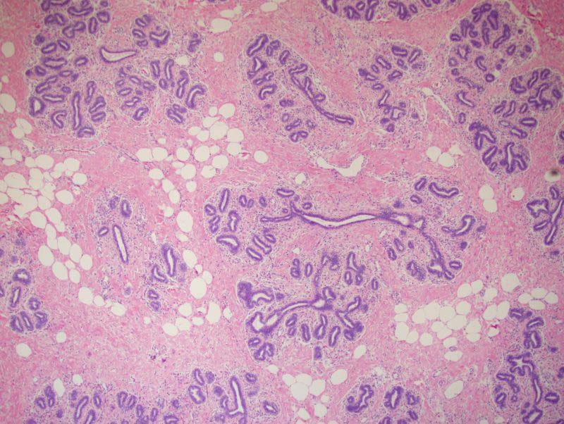
Histologically, the mass demonstrated dense, eosinophilic fibrous tissue surrounding ductal and lobular structures that form intact terminal ductal lobular units (Figure 1, 10x). The epithelium consists of luminal and myoepithelial cells without atypia or hyperplasia noted (Figure 2, 40x). Located between the ductal lobular units, there are scattered adipocytes mixed with fibrous stroma (Figure 3, 10x) and larger ductal structures that have an angular appearance.