Case of the Month: Pediatric Gastric Mass
By Roula Katerji, MD, PGY-2
Clinical History
An 11-year-old female presented to the ED with five episodes of coffee ground emesis, mild abdominal pain and pale mucosal membrane. She was overall awake, alert and interactive.
Past Medical History
The patient was previously healthy with no clinical problems.
Recent History
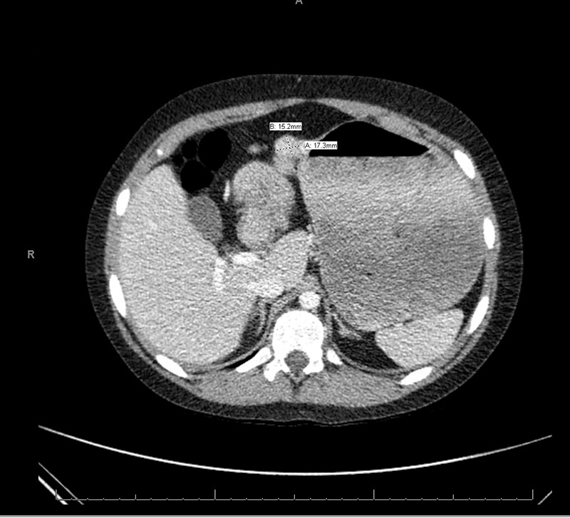
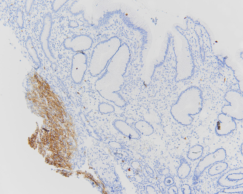
Initial laboratory work up demonstrated that the patient was anemic (Hgb = 4.5 g/dL), with WBC and platelet count within normal limits. CT scan revealed two exophytic masses originating from the greater curvature of the stomach at the antrum with intra and extraluminal components (Figure 1). Upper endoscopy noted a few small, smooth sessile polyps in the gastric antrum. One large ulcerated polyp was noted close to the pylorus.
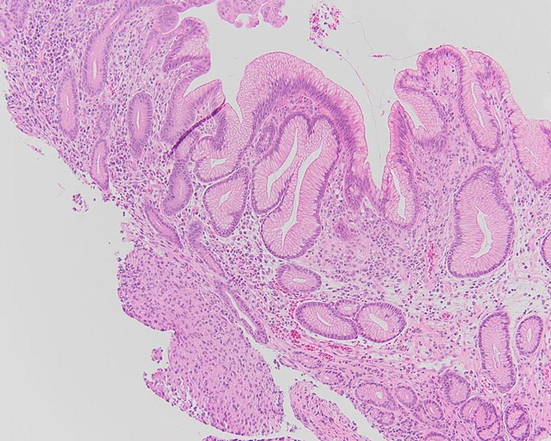
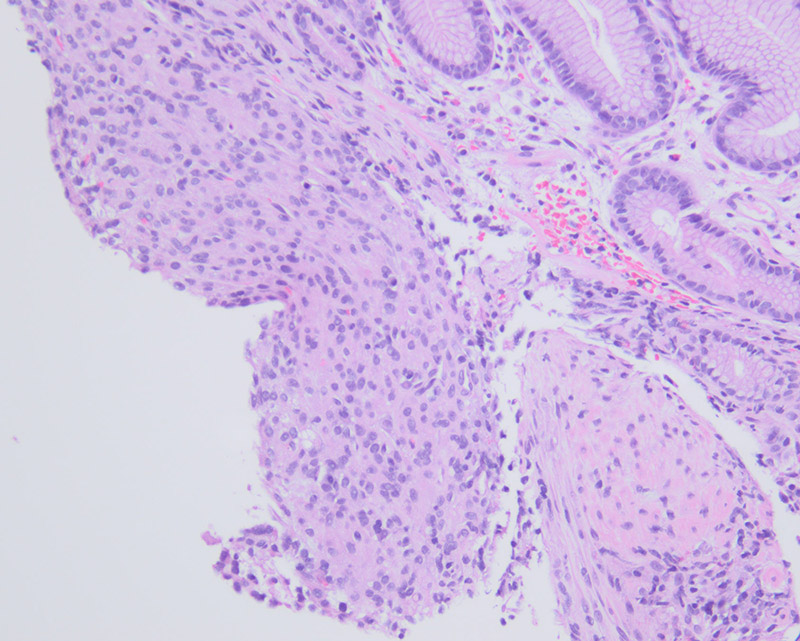
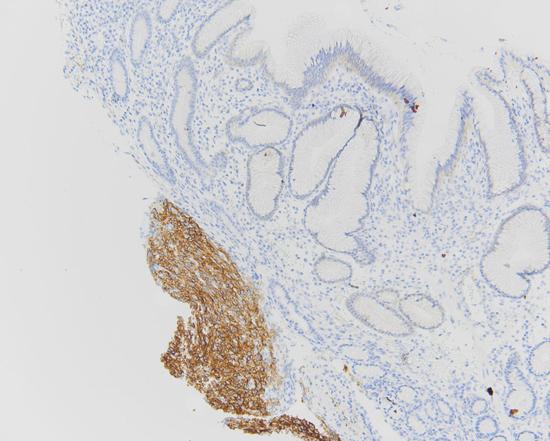
The polyp was non-obstructing and not actively bleeding; biopsies were taken of this polyp. Histologically the biopsy specimen demonstrated a proliferation of uniform mixed epithelioid and spindle-epithelioid cells amongst antral-type mucosa (Figures 2, 3). No significant pleomorphism or mitotic figures were identified. Immunohistochemical stains for DOG1 and c-Kit (CD117) were positive (Figures 4 and 5, respectively). A subsequent wedge resection of the stomach to include the lesions was performed and confirmed the original biopsy diagnosis (Figure 6). Molecular testing for c-Kit and PDGFRA were performed and were negative.