URMC / Labs / Zhong Lab / Projects / Clinical Diffusion Imaging
Clinical Diffusion Imaging
As an example, in the work of Bazarian et al (2007) we show that collectively, DTI detected significantly lower trace and elevated FA values in mild TBI subjects compared to controls. These abnormalities correlated to poor clinical outcome. We believe these findings represent axonal swelling, an early step in the process of axonal injury. Furthermore, we investigated the ability of wild bootstrapping analysis to detect subject-specific changes in brain white matter (WM) before and after sports-related concussion (Bazarian et al 2012). Wild bootstrap analysis detected significantly changed WM in a single concussed athlete. Athletes with multiple subconcussive head blows had significant changes in a percentage of their WM that was over three times higher than controls.
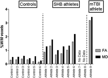 Total change ratios in FA and MD for each subject. The number
of voxels with significant preseason to postinjury/season changes
in FA and MD is quantified by the percentage of all WM voxels
with a significant change (either increased or decreased)
Total change ratios in FA and MD for each subject. The number
of voxels with significant preseason to postinjury/season changes
in FA and MD is quantified by the percentage of all WM voxels
with a significant change (either increased or decreased)
in FA or MD. Change defined as (postseason)-(preseason).
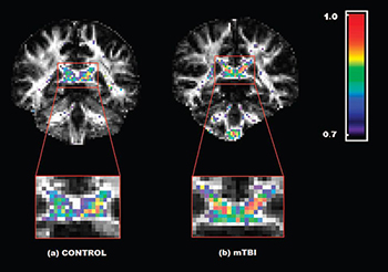
Fractional anisotropy (FA) values in the posterior corpus callosum
of a selected control and mild traumatic brain injury (TBI) subject.
Color-coded FA values in the posterior corpus callosum of a
selected control subject (left) and a mild TBI subject (right). The
color scheme is NIH color space; higher FA values are red, and
lower ones are blue. The color threshold is set to FA _ 0.7–1.0.
Note that the mild TBI subject has more red voxels (higher
FA) and the control more blue voxels (lower FA).
« back to all projects