menu

March 2016

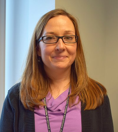
URMC Representative Lays the Groundwork for Pathology to Combat Cancer in Haiti

3/28/2016

ayooo

Vendors sell goods in the city of Cape Haitien, Haiti, which is the poorest nation in the Western Hemisphere.
 

Loralee McMahon was shocked as she asked a Haitian ob-gyn doctor and nurse how surgical specimens were used after an operation.

McMahon, 41, is the supervisor of Immunohistochemistry (IHC) in the Surgical Pathology text division at the University of Rochester Medical Center.

In February, she took a solo trip to Haiti where she spent two weeks volunteering in the nation's two largest cities. Her task was to set up a full working histology lab at a hospital and train local lab technicians in IHC.   

As she spoke with two women whom she was staying with in the city of Cape Haitien, McMahon was about to get a reality check.

"I said, I'm trying to train people in histology and it would be great if you guys could take the specimens to the lab. What are you doing with the specimens now?"

"We just throw them away," they responded. 

Haiti is the poorest country in the Western Hemisphere and, with over 10 million people, the second most densely populated. Like most low income countries, the state of healthcare is bleak and options for cancer diagnosis and treatment are few. 

Enter McMahon, who was first inspired to help after hearing Dr. Jennifer J. Griggs speak at URMC. Griggs is a medical oncologist based at the University of Michigan who previously completed a postdoctoral fellowship at UR. 

Griggs took a one-year sabbatical to work with two schools with nursing programs in Haiti, which, like many low income nations, lacks the heath care infrastructure to diagnose and treat cancer.

In addition to poverty and overcrowding, Haitians face many barriers to treatment, especially when it comes to breast cancer. Many women live with symptoms of breast cancer before seeking medical attention, Griggs explained. As in other countries, they often fear what a diagnosis might mean for their lives. 

"(Women fear) what treatment will involve, losing a breast and no longer being desirable if they lose a breast,” Griggs wrote in an email. “They fear that, even if they get treated, they will still ultimately die of their disease. Major public health efforts are needed to inform women that medical care, when delivered early, can improve their chance of cure.”

After her presentation, Griggs asked how to set up a pathology lab for breast cancer specimens. McMahon and other staff put together a list of equipment to summarize the supplies needed to build a histology lab from scratch. McMahon's interest in joining the effort started to snowball from there.  

She contacted Dr. Vincent DeGennaro Jr., an internal medicine doctor and public health specialist at the University of Florida who works for the nonprofit Project Medishare, an organization that built a bbbbbhospital in the capitol city of Port-au-Prince after the devastating 2010 earthquake rocked Haiti. 

After postponing the trip due to political unrest in the country, she arrived in February 2016. McMahon spent the first week of her trip in Port-au-Prince where she trained two young lab technicians at the National Public Health Laboratory (NPHL) and began to set up immunochemistry (IHC) testing on site.

Right: Members of a Project Medishare breast cancer support group (photo courtesy of Vincent DeGennaro).
 

IHC – the detection of proteins indicative of certain diseases in tissue cells – is almost nonexistent in Haiti. Pathologists are extremely rare, and work in private pathology labs that lack resources to perform IHC. As a result, many send their specimen slides to the U.S. for IHC testing. The resulting turnaround time for lab results is outrageous and the need for independent testing greater than ever.

The mindset of healthcare providers in Haiti is different from that of Western institutions. Hospitals are overcrowded, so treatment must be quick. If a patient has surgery to remove a tumor, the problem is considered "fixed" unless the lump comes back. The tissue is thrown away and the patient is sent home. Needless to say, these conditions put medical research at a great disadvantage.

During the second week of the trip, McMahon saw just how severe things were on the ground. Gone was the air-conditioning and running water at the NPHL in Port-au-Prince as she and her trainees went to Justinian Hospital in the city of Cape Haitien. 

The hospital was a collection of fenced-in buildings guarded by armed security while sick and injured patients and their families crowded into an outdoor waiting area. On the first day, McMahon turned on a faucet to wash her hands and nothing came out, so she started bringing jugs of water to work each morning.

cccThe outdoor waiting area at  Justinian Hospital. Below: The early beginnings of a histology lab.


Despite the difficult working conditions, the two trainees showed great enthusiasm as McMahon instructed them via the translator who was hired off the street. They were laying the groundwork for a working histology lab at the hospital. They were making small advances every day as McMahon learned to work through interruptions, like frequent power outages that brought her lessons to a halt.

"There were a couple times during the week where I was like, this is way too much," she said.  
"I'd come home at night and be like, this is just pointless - Why am I here? Then the next morning the techs would be there before I arrived and be excited and ready to learn."

It's the start of real progress, and McMahon hopes momentum will continue to build as more volunteers provide help. At the time of this interview, a breast pathologist from Canada planned to go in March and bring lab supplies to Justinian Hospital.  

Four additional volunteers were slated to go as well, but the project was stalled because the Haitian government would not renew expired paperwork authorizing the crew's large equipment to be taken into the country.  

When the lab equipment is finally installed, McMahon hopes the lab at Justinian Hospital will start to see specimens trickle in. As Haitian doctors and nurses start to realize the benefits of pathology in patient treatment, she believes the nation has a fighting chance to combat the cancer epidemic. 

"I started telling the surgeons to save their (tissue) specimens. Even if you got a lab tech to take the specimen at least get is started so it doesn't go to waste," she said. "At least practice." 

McMahon says she eventually wants to go back to help move the mission forward, but understands that real progress will not happen overnight.  
 
"By the end of the two weeks I saw that this is way bigger than making a histology lab," she said. "But you have to start somewhere."

For information Project Medishare, or to make a donation, click here

Pathology Welcomes 2016 Resident Class

3/21/2016

The Department of Pathology and Laboratory Medicine is pleased to welcome four new residents in July 2016. The incoming class includes:  

ALT


Alexandra Danakas Lake Erie College of Osteopathic Medicine 

ATG


Roula Katerji University of Aleppo Faculty of Medicine

NOOR


Mushal Noor - King Edward Medical University 

REYES


Cynthia Reyes-Barron - Texas Tech University Health Sciences Center Paul L. Foster School of Medicine 

Returning residents Shana Stroub and Sachica Cheris will serve as the Chief Resident and Associate Chief Resident respectively.

stroub


Shana Straub, M.D.

cheris


​Sachica Cheris, M.D.


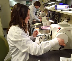
Not Just Cut and Dry: A Day in the Life of a Histologist

3/7/2016

histo1
Behind many patient diagnoses is a process that starts in the operating room and ends under a microscope.  

UR Medicine Labs has a team of histotechnicians (HT) and histotechnologists (HLT), often referred to as "histotechs," who work around the clock to complete the steps leading up to patient diagnoses for cancer and other diseases.  
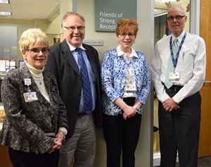
Histotechs are responsible for taking human tissue to prep, cut, and stain before it's examined by a pathologist. We recognize this important work in honor of Histotechnology Professionals' Day on Thursday, March 10. 

"I think most people have never heard of histology," said histotech Wally Pryjmak, who has worked at UR Medicine Labs for two years. He didn't even know what a histologist was when he became a certified medical lab technician. Nevertheless, Pryjmak wants patients to understand the link between his work and their treatment. As he says, "We create the slides your diagnosis is based on, so without us it would be a lot harder to get diagnosed." 

The tissue prep process starts after a patient has surgery to remove tissue from the body, often in a biopsy. This tissue is brought to the Surgical Pathology area (located on the ground floor of SMH) and is most often examined first by a pathologist's assistant (PA) who dissects the tissue at a grossing station.  

The tissue sample then goes into the hands of a histologist who processes it through a series of alcohols to remove any water, clears tissue in xylene and infiltrates it in paraffin wax. The tissue is then embedded into a paraffin block and the histologist cuts it into paper-thin slices.

histo2Wax slices of tissue then go into a warm water bath to stretch them out and prevent wrinkles before carefully placing the tissue onto a glass microscope slide. The histologist gently scoops the section of tissue onto the slide to dry. He or she then treats the slide with a basic stain (hematoxylin and eosin) to make the sample is easy to see under the lens. 

There are up to 75 different stains that reveal certain tissue elements, fungus, or bacteria. A pathologist can request any combination of stains be added before viewing the slide close-up. 

In this way, the histologist is part artist and part chef: they must follow a "recipe" of stains to yield a proper reading. Even a small misstep can compromise the diagnosis. Once the slide is treated with the proper stains, it goes to a pathologist who examines it and issues the diagnosis.

The work of a histologist requires careful attention to detail and physical stamina. On average, the histology team at Strong prepares between 800 and 1,200 slides each day.

histo3Diana Scott is the supervisor for histopathology at Strong. To her, the work of a histologist is like an art form. And while patients don't see or speak with the lab team helping behind the scenes, the connection is still very meaningful. 

"We care about the production side of things and the quality of the slides," said Scott. "We treat every specimen like it belongs to one of our family members waiting for their test results to come back." 

In photos:

(Top) Histologist Lisa Torregrossa embeds a tissue sample into paraffin wax. Once the wax hardens, the block can be sliced for examination. (Top right) Elizabeth Pilon uses a microtome instrument to slice a piece of human breast tissue that has been embedded in wax. 

histo4(Above right) There are more than 75 special stains used to dye tissues so they can be viewed clearly under a microscope. The stains highlight specific tissue components (nuclei, muscle, etc.) or microorganisms (bacteria, fungus) and a combination of stains is often necessary.

(Right) Tissue blocks, or cassettes, are preserved and filed for at least 20 years after a diagnosis is given. 


Pathology resident room gets a facelift

3/1/2016

resroom1In February, Pathology residents celebrated the opening of a new and improved study room.

Renovations to the space included new cubicle walls, carpet and a fresh paint job.

The room, which serves as a meeting place and quiet study area, is located on the second floor at 2-6440.

It was previously home to cytopathology, whose office moved to 2-2100 in 1997.

resroom2In photos: (Above) Pathology residents Shira Winters, Nisha Patel, and Hani Katerji in the newly renovated resident study room. (Bottom) Sohaib Abu-Farsakh examines slides.

Pathology plays key role in tissue bank partnership

3/1/2016

IndivumedIn photo: From left, Dr. David Linehan, chair of Surgery for the School of Medicine and Dentistry, Dr. Hartmut Juhl Founder and CEO of Indivumed, Dr. David Hicks, Dr. Bruce Smoller, and Andrew Deubler, deputy vice president for University Advancement.

About the partnership
 

The department of Pathology and Laboratory Medicine is now the gatekeeper of a human tissue bank at the University of Rochester Medical Center (URMC).

URMC has signed a 3-year contract with German-based company, Indivumed, to allow patients to give access to having their tissues classified and used for cancer research worldwide.

The operation is fully funded by Indivumed, a startup founded by Dr. Harmut Juhl in 2002. The company will fully fund the biobank in exchange for half of the available samples.

How does it work?
 

Patients at Wilmot Cancer Center will have the option of giving consent to allow samples of their tissues - taken from biopsies and other procedures - catalogued and used for research. When consenting patients are in surgery, an employee will be stationed in the operating room to immediately take tissue to Surgical Pathology to be examined by a pathologist.

The process must be completed within moments of the extraction. When the tissue arrives, a pathologist must first examine a segment to determine a patient diagnosis. Once confirmed, the remaining tissue is divided into samples that are labeled and entered into a database. Some will be set aside for Indivumed's use.

Dr. David Zhou serves as director of the bank. Dr. David Hicks helped establish protocol for managing samples from the second they arrive to Surg-Path.

How will it be used?
 

URMC has not yet established protocol for deciding who gets access to the tissue first. Dr. Bruce Smoller, Pathology chair, said protocol will be determined by a group of URMC leaders.

"(Participating) will increase our profile in the research realm at URMC," said Smoller. "One of the goals is to build our research profile by accessing the available resources."

You may also like

No related posts found.