
1/16/2017

Name: Krisztina Hanley, M.D.
Hometown: Originally from Hungary and now lives in Decatur, Georgia
Family: Husband, Jim, and daughters, Aideen, 6, and Maeve, 3
Occupation: Assistant professor of Pathology & Laboratory Medicine, medical director of macroscopy and gross rooms, Emory University Hospital and Emory University Hospital at Midtown, rotation director of Gynecologic Pathology, Emory University Hospital.
Education: M.D. from University of Pecs in Hungary (2001), AP/CP residency at University of Rochester Medical Center (2003-2007), Cytopathology Fellow; Emory University School of Medicine (2007-2008), Gynecologic Pathology Fellow; University of Virginia (2008-2009).
Current research: A phase II clinical trial examining an agent that targets folate receptor alpha. She is also working with residents on a project that’s looking at certain pathways that might be connected to a certain pattern of invasion in endometrial cancer. Finally, she is researching a marker called OTP that can distinguish pulmonary versus non-pulmonary well-differentiated neuroendocrine tumors.
What first brought you to the U.S?
My husband is from Rochester and we met when I was still in medical school (in Hungary). It was a long-distance relationship. We wanted to live in same city so that’s what brought me to Rochester. We got married when I was a first-year resident in 2003.
Now you work at Emory in Atlanta, GA, where you completed a fellowship after residency. What aspects of that program made it attractive to you?
At Emory, the cytology fellowship is unique because we see a lot of patients. We have a clinic where we do fine needle aspirations. We perform the FNA ourselves, look at it, and talk to the patient. Sometimes we tell them the results right away. It’s very unique that as pathologist we actually see patients.
A lot of people think pathologists just sit in their office and look at slides or do autopsies in the basement. We actually do see patients. Also, the FNA clinic is in the cancer center (Winship Cancer Institute) which gives us the chance to have a very close relationship with the clinicians, the oncologists.
When you reflect on your time as a resident, what do you think has prepared you most for your career?
In retrospect, there are things I am glad I did even though I may not have liked them at the time. For example, in the gross room we were really busy with gross specimens. I remember very heavy days that made me miserable! I’m glad because even now after many years, I can still handle most of the specimens without any problems. I became very efficient. That’s also true with autopsies. We had a lot of autopsies at UR compared to other programs, and I think that really helped me get through things even when it gets really busy. I don’t freak out anymore.
For frozen sections it was the same way. We used to cut our own sections at UR. At Emory, they have a lot of help from PAs. I’ve been out of training for seven years now and I don’t have a problem cutting a frozen section. This is huge because when residents aren’t available or we have multiple frozen sections in the same time, I can help out.
Did you have any mentors during your time at URMC?
A few people had a major impact on me and the way I approach things: Dr. Thomas Bonfiglio who is retired, Dr. Ellen Giampoli, and Dr. James Powers. From a clinical pathology (CP) side, there was Dr. Marilyn Menegus, Dr. Neil Blumberg, and Nedda Howk from the Blood Bank. I don’t do hematopathology but I can thank (the late) Dr. Ray Felgar and Dr. Arnaldo Arbini for everything I know in hematopathology. The cytotechnologists at UR are outstanding. They are very engaged in resident education. Michael Facik, Donna Russell, and Mary Ann Rutkowski had major impact in my training in cytopathology and fellowship choice.
How do you like to spend your free time?
I love to bake. I cut back on that because my husband and I end up having to eat everything or it goes to waste, since my kids may or may not like what I bake. I started running after my older daughter was born. In Georgia you can run or hike outdoors pretty much the whole year, so we have hiking sticks for the girls and try to spend as much time outdoors as we can.
What do you think it’s going to take to draw more young people to the field of pathology?
I think they need to get early exposure. Exposure in medical school is very limited and there’s a lack of understanding for what pathologists actually do. So, we need to reach out to medical students and allow them to have hand on experience in our department. This could be sign out, gross room activities, participate in frozen sections, attend tumor boards and come to FNA clinic. Most people, including physicians from other specialties, have no idea how diverse and complex the work of a pathologist is.
1/4/2017
Pregnancy can be an anxious time for women, especially those who may be at higher risk for certain complications.
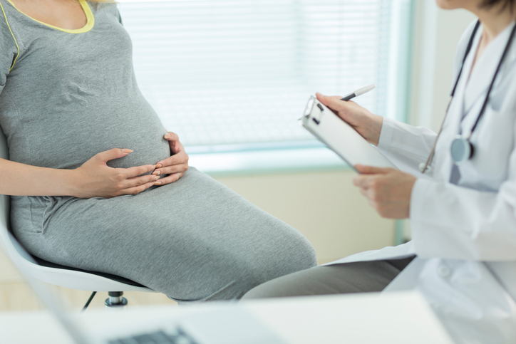 UR Medicine offers prenatal testing for expecting mothers at the Biochemical Genetics Laboratory at Strong Memorial Hospital.
Together with Department of Obstetrics and Gynecology, this team of clinical professionals helps provide families with answers to crucial questions before a baby is born.
How it Works
In New York State, all pregnant women in their first trimester are offered an optional blood test to screen for certain fetal defects. The patient’s OBGYN can order the test and have her sample sent to the laboratory.
From there, a team of medical technologists process the sample on an automated lab instrument that issues a report with the odds of certain complications. These results go to the ordering physician within 24 hours.
The first trimester maternal serum screening test assesses the risk for chromosomal defects – Down syndrome and Trisomy 18 – by combining the blood test result with a specific ultrasound measurement. Expecting mothers can later have a second trimester AFP only screening for spina bifida.
If the first screen comes back negative, no further testing is required. If the result is positive, patients are directed to a prenatal counselor who meets with them to take a more in-depth look.
The risk level depends on a number of factors including age, ethnicity and family history. While the first trimester screening provides an assessment of the risk, it is still too early to determine a diagnosis.

Jeanne Peterson
Jeanne Peterson is a reproductive genetics counselor at URMC who has spent more than 30 years consulting families facing possible or likely issues during pregnancy.
The vast majority of positive screens in the first trimester are actually false positives, she explains. Still, parents are often alarmed when they hear there is a higher-than-average risk of something going wrong.
“Once the patient gets that phone call from their doctor that they weren’t expecting, this cloud comes over them,” said Peterson. “Many times they’re not getting all the information about the results, and even the information they’re getting they might not understand very well.”
For example, an expecting mother with a 1 in 200 chance (0.5 percent) of having a baby with a birth defect is at much lower risk than someone with a 1 in 6 chance. By learning about their test results and what the numbers mean, many walk out the door feeling reassured and prepared for next steps, whatever they may be.
“You have to help people through it,” said Peterson. “You have to empower them by giving them information at the level they can understand.”
Higher risk individuals with a positive first screen can decide whether to have optional follow up testing for chromosomal abnormalities.
Advances in the last decade have provided a less invasive option than the traditional procedures, explains Dr. Robert Mooney, director of the Biochemical Genetics Laboratory at URMC.
Patients used to be limited to amniocentesis – a test that samples the amniotic fluid around the fetus – or CVS – which requires a small sample of the placenta early in pregnancy – to get a definitive diagnosis. Today, there is a less invasive option that often eliminates the need for more invasive procedures.
Robert Mooney, Ph.D.
The cell-free fetal DNA blood test is available for mothers as early as 10 weeks of gestation. This test analyzes genetic material from the placenta that is present in a woman's blood during pregnancy. It can accurately eliminate most false positives and identify those pregnancies with a high risk of an abnormality.
“This has really taken over as the next step after we identify a screen positive,” said Dr. Mooney. “In most cases this eliminates the false positives and identifies those who are at very high risk. We’ve now narrowed the population down to a few at very high risk rather than 3 to 5 percent who have screened positive by our (first trimester) blood test.”
Patients who still test positive after the cell-free DNA test can then choose to have amniocentesis or CVS to obtain a definitive diagnosis.
A Team Effort
The prenatal screening program at URMC is a combined effort of the Department of Obstetrics and Gynecology and the Biochemical Genetics Laboratory. Representatives from both areas meet regularly to review individual cases, changes to testing, or population trends.
“We’re part of a team,” said Dr. Mooney. “Prenatal Screening is successful only because we all work together. We communicate (with OBGYN) constantly and they give us information to help us interpret the results appropriately.”
The lab considers each test to represent a person and a family waiting anxiously for answers. The team operates on the presumption that every single result is important, says lab supervisor, Matthew Morriss.
“Each sample is unique,” he said. “It has a person at the end of it and we treat each one with the same urgency. These results are important and we want the doctor and the patient to have all the information that we can give them.”
As a counselor, Peterson says she finds it rewarding to help expecting parents be better prepared for the next step of their journey.
“I tell them that most babies born to all couples are healthy and normal and most likely this baby is healthy and normal, too,” said Peterson. “These screening tests sometimes create bumps in the road but the majority of the times, things turn out okay.”
12/15/2016
They say the best way to learn is to teach, and for the first time ever, licensed laboratory technologists at URMC will do just that through a new clinical laboratory technology program.
 The program will provide full-time clinical lab education for prospective medical technologists, with lectures and hands-on clinical training leading to an advanced certificate. The University and the New York State Education Department have approved the program, and it will welcome its first class of students in fall 2017.
Applicants must have a bachelor’s degree in the biological, chemical or physical sciences and have completed the coursework required for state licensure.
The University had previously partnered with Rochester Regional Health System (RRHS) to provide clinical training to students who received the lecture and exam portion of their training at Rochester General Hospital (RGH), but will now provide both facets of training on its own.
Vicki Roberts, program director and manager of education for the Department of Pathology and Laboratory Medicine, says the region needs every training program working at full capacity to fill a growing number of vacancies in the field.
“This is a benefit to the University and the region because it gives people who are unable to find a practical application for their degree entry into a licensed professional position,” Vicki says.
In 2006, New York State changed its licensing requirements for medical technologists (“med techs” or MTs). This law meant that staff who previously needed a B.S. degree in an applicable major must now complete 1-2 years’ worth of additional clinical training and pass a certification exam in order to be state-licensed.
While many MTs were “grandfathered” in when the law changed, others have balked at the new, more demanding educational requirements. This has made it more challenging than ever for employers to fill vacancies in the lab.
Leadership’s hope is that this new training program allows UR Medicine Labs to have a steady pipeline of trained, certified technologists to fill these vacancies as we grow and affiliate with more partners throughout the region—from Strong Memorial Hospital (SMH), Strong West and Highland hospitals, to medical campuses at FF Thompson in Canandaigua, Dansville, Wellsville and Hornell.
“UR Medicine’s need for additional licensed medical technologists could not be more urgent,” says Kathy Parrinello, chief operating officer of SMH. “This training program allows us to bring in current and prospective medical technologists to train in our excellent labs at SMH, graduate, and get their licenses so we can hire them into positions,” she adds. “We are grateful to Vicki and the entire team for their diligence and perseverance in bringing this program to fruition.”
The majority of lab staff at URMC is comprised of licensed MTs that work around the clock to perform a range of diagnostic tests. These tests help doctors learn what’s making patients sick and properly diagnose and treat them.
Med techs work in labs including Blood Bank and Transfusion Medicine, Microbiology, Chemistry and Hematology, Flow Cytometry and Bone Marrow Testing, Molecular Diagnostics, Surgical Pathology and more.
 Geoffrey Harris (right) spent the last four years as Education Coordinator in the Hematology Lab. He’s one of many MTs that will serve as instructors in the new program.
“When everyone in a lab is an instructor and everyone teaches, it keeps people on their game,” Geoffrey says. “You realize this is a good thing for the whole lab and I think it makes everyone stronger.”
The new class will have between eight and twelve trainees who must complete 35 credits of non-clinical work and 720 hours of clinical experience before taking their certification exam.
 People like Caroline Brown (right) know what it’s like to have a long path to licensure. She works in Clinical Microbiology, which is one of the largest labs at SMH—in terms of staffing and number of specimens.
When she started as a med tech at URMC, she simply had a B.S. degree. She took time off for family reasons and soon found that returning to work was not as easy as she’d hoped.
“In that timeframe, the licensing all came into being and I fell through the cracks,” Caroline says. “I had to do something in order to get back into the lab.”
She was accepted into the RRHS training program, which she completed, and later returned to UR as a licensed MT. Today she teaches trainees like herself who are hoping to grow their careers.
Teaching means MTs have new responsibilities on top of their regular workload, taking extra time and preparation to educate students.
For Caroline, that means strategically preparing live cultures days in advance so that students are able to simulate the work that licensed techs perform on a daily basis. This kind of prep is critical in making students’ experiences as authentic as possible so they are prepared to work in a lab.
Caroline says playing a part in this instruction is the best way to give back so others can have the same opportunity she did. “I feel for the future of the career in the lab,” she says. “We need people who want to learn and want to be here.”
The Medical Technologist program is now accepting online applications. For questions, contact Vicki Roberts at (585) 276-3688 or Vicki_Roberts@URMC.Rochester.edu.
12/6/2016
 The American Society of Cytopathology (ASC) has recognized two individuals linked to the Department of Pathology and Lab Medicine at URMC.
Kathryn Kiely (top, right) was one of five cytotechnologists from across the U.S. selected to receive a travel scholarship to attend the ASC's Annual Scientific Meeting to be held in Phoenix, AZ in November 2017.
Kelsey Snyder, (below, right) the first student to graduate from the Roswell Park Cancer Institute/Daemen College cytopathology training program, where Donna K. Russell, M.Ed, CT (ASCP) HT is the program director, also received a travel scholarship to the ASC meeting.
 Snyder was additionally named a recipient of the 2016 Geraldine Colby Zeiler Award, which is given to five cytotechnology students who show great microscopic diagnostic skill, leadership and initiative within their program.
10/13/2016
 If you’ve ever had a sore throat swabbed to test for strep, you have experienced just one way bacteria cultures can be used to help sick patients get answers.
The Bacteriology Laboratory at Strong Memorial Hospital runs hundreds of tests around the clock to identify the bacteria and fungi that cause everything from urinary tract infections to food poisoning. This identification process is the first step in stopping sickness in its tracks and putting patients on the road to recovery.
There is a rainbow array of plates that medical technologists use to grow different bacteria. An ordering provider may suspect a certain type of infection and ask the lab to run a specific test, or tests, to confirm the identity of the culprit and see what drugs it best responds to.
Most bacteria are traditionally grown on an agar media plates that contain sheep’s blood. Historically, this has been the way to obtain bacterial growth in order to determine which organisms are “normal flora” and which bacteria are “bad,” causing disease or infection. Culturing allows “bad” bacteria to be tested for susceptibility to certain antibiotics. Advances in modern technology are now making this process more automated than ever before.
How it starts
If you have symptoms of a urinary tract infection, for example, your urine sample will be sent to the lab for testing from your doctor’s office. After it has been received, labeled, and entered into an electronic database for tracking, the specimen goes to a medical technologist.
He or she will then dip a tiny calibrated plastic loop into the urine and make a streak onto a sterile blood plate to start the process of growing bacteria. This process is called inoculation.
About 80 percent of all UTIs are caused by the bacteria E. coli, classified as Gram-negative bacilli due to the composition of the cell wall. The tech will also select media (another name for agar plates) that will only grow Gram-negative bacteria (called MacConkey agar in this case) to help identify the “bad” bacteria and prevent growth of other organisms.

“That’s what a lot of bacteriology is: knowing who the good guys are – the normal flora – and who the pathogens are,” said Jennifer Barrante, education coordinator for Microbiology at URMC. “We’re trying to enhance the recovery of our ‘bad’ guys.”
Since bacteria need warmth and time to grow, the inoculated plates are then placed into and incubator that looks like a refrigerator with a glass door, but warms up to body temperature instead of cooling. The plates are allowed to incubate for a minimum of 18 hours to give the bacteria time to grow.
After incubation, the technologist takes the culture and counts the number of bacterial “colonies” that have grown overnight. E. coli, for example, will appear as pink colonies on the MacConkey plate after it has incubated overnight. This is a quantitative culture because the quantity of bacteria reported is based on the actual number of colonies grown, multiplied by 1,000 to determine colonies per milliliter of urine.
Most cultures are not quantified this way. Instead of counting the number of colonies, cultures are usually read semi-quantitatively using a four-quadrant system.
Picture the circular plate evenly divided into four sections with a specimen streak in each quadrant. If growth appears only in the first quadrant where the specimen was inoculated, the reading is “one-plus.” If there is also growth in the second section of streaking, it’s 2+, and so on.
What else can you grow?
Lab techs are able to culture bacteria from a wide variety of biological material. This can be a swab of something as innocuous as a blister or a cut on your finger, to something as serious as spinal fluid to detect meningitis.
If a patient undergoes a biopsy on a tissue mass that’s suspected to be cancerous, part of the tissue specimen can be cultured to see if the growth is caused by an infection instead of cancer.

Blood specimens can also be cultured to detect bacteremia (bacteria in the blood) or sepsis (a severe reaction to an infection that may involve the blood) which can be caused by different organisms like staph, or Haemophilus.
Besides urine and blood, stool specimens are routinely cultured to identify bacteria – like Salmonella, Shigella, or Campylobacter – that cause food poisoning.
You can also test for fungal infections like ringworm, a yeast infection or oral thrush, which appears as white spots inside the mouth.
The lab also tests specimens for acid-fast bacilli, a group of bacteria that includes the cause of tuberculosis.
Automation
While these methods of culturing specimens have been around for many decades, new technologies allow labs to get the same results more quickly, and with less hands-on work.
There are several large instruments at URMC that perform testing on a molecular level. This kind of test is called a PCR, or polymerase chain reaction, and it uses small samples of DNA to detect patterns that indicate the presence of certain bacteria, viruses, or infections.
 One PCR instrument can test stool samples for presence of C. difficile and Norovirus. That same instrument can use a nasal swab to test for MRSA. Another instrument allows for testing of the respiratory virus panel based on a nasal swab.
The newest instrument, the BD MAX, has replaced the traditional stool culture method for detecting Salmonella, Shigella and Campylobacter. It can also detect the stool parasites Giardia, Cryptosporidium, and E. histolytica when requested by a doctor.
This instrument reduces the time it takes the result to get to the doctor. A traditional stool culture can take 3-4 days to be resulted once received in the lab, but the BD MAX can get the same results within 24 hours.
Automated molecular instruments can also provide much faster results for some sexually transmitted diseases just hours after they are run on the instrument, rather than patients having to wait days for traditional cultures to yield a result.
Fighting back
Yes, PCR automation saves time, but identifying the causative agent of an illness is just one step in determining how it should be treated. Many specimens must still be cultured to determine their susceptibility – to find out which antibiotics will kill off the offending bacteria. This process is called susceptibility testing.
 One method of S.T. is the Kirby-Bauer disk diffusion test. For this test, the surface of a large culture plate is swabbed with the pathogenic organism and small discs of antibiotics are placed on the plate. After incubating overnight, a tech will measure the diameter of the circles that have formed around each “pill” to determine what medications are most effective.
"We have different panels of drugs to test depending on the bacteria and specimen source the organism comes from in order to provide the best treatment options,” said Barrante.
She explained that using antibiotics incorrectly can wipe out populations of very susceptible bacteria and can leave more resistant strains behind. Or, organisms can swap resistance genes and cause “superbugs” that can’t be killed by antibiotics.
Susceptibility testing informs the clinician of the antibiotics that can be used, as well as those drugs that the bacteria is resistant to. This provides a range of responses to determine the most effective treatment, even if it means testing more than one drug.
One person’s story
With thousands of lab tests making their way through the Bacteriology Lab each month, it’s important to remember that each one represents a patient who needs help.
Barrante recalls her own experience with severe illness as a child, and how it inspired her to get into the field of Microbiology.
She remembers the discomfort of getting her blood drawn frequently, and feeling like a “human pin cushion.”
Since being treated, Barrante says she has always wanted to give back, and becoming a medical technologist was the way to do it; performing tests that guide the doctors who treat patients.
“I wanted to help people because I was sick and somebody in a lab somewhere figured out what was wrong with me,” she said.
In photos
Top: A medical technologist inoculates an agar plate with a small amount (approximately 1 microliter) of urine. The plate will incubate in a warm environment to allow bacteria to grow.
Second from top: E. coli bacteria shown in this culture is consistent with what causes most urinary tract infections.
Third: A Haemophilus bacterial infection is visible in the lower plate, which is a media called chocolate agar due to its color.
Second from bottom: Medical technologist Marie Rouse holds a respiratory virus panel, a multiplex PCR test that will run through an automated testing instrument.
Bottom: The Kirby-Bauer disk diffusion test determines what antibiotics are most effective in fighting the bacteria that has grown in culture.
You may also like